Fondamenti Scientifici (hub)

Dalle scienze fondamentali alla traslazione clinica

fundamental-sciences divider-fundamental

Schema Sperimentale

einstein. divider-fundamental
  • istologia

    Istologia

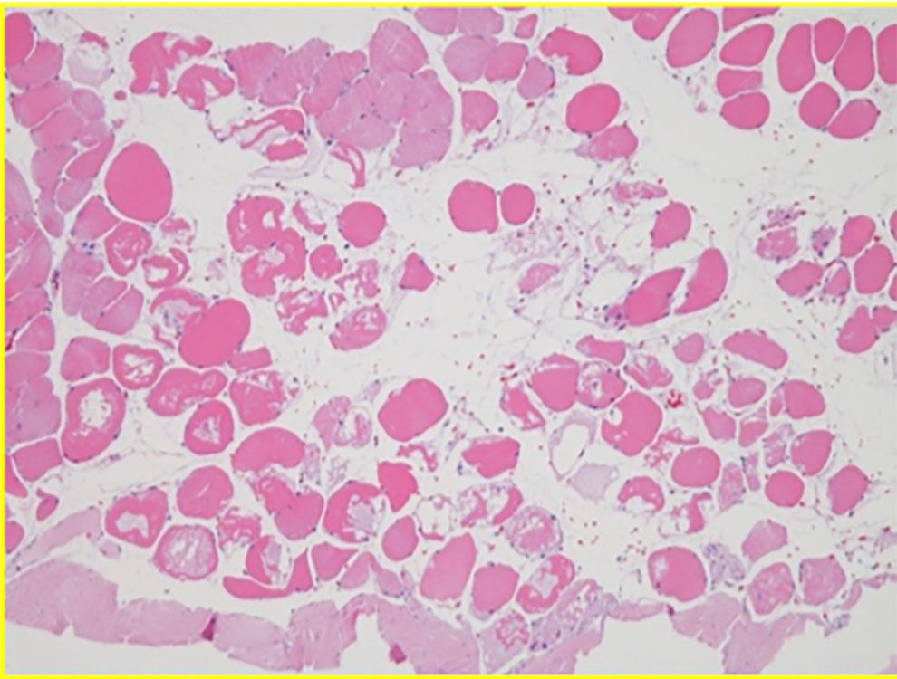
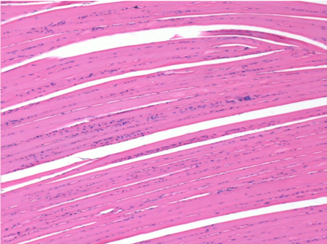
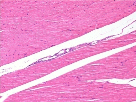
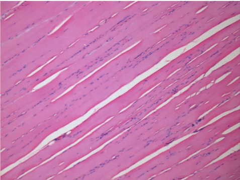
    Sezioni istologiche • Architettura cellulare • Risposta tissutale

    View Histology

  • chemistry

    Chimica

    Modulazione del pKa • Composti aromatici • Stabilità chimica

    View Chemistry

  • mecanism-action

    Meccanismo d'azione

    Risposta biologica integrata • Vie metaboliche

    View Mechanism of Action

  • molecular-biology

    Biologia Molecolare

    Segnalazione intracellulare • Regolazione molecolare

    View Molecular Biology

  • physiology

    Fisiologia

    Fisiologia neuromuscolare • Anatomia funzionale

    View Physiology

  • lab of anatomy

    Studio cadaverico

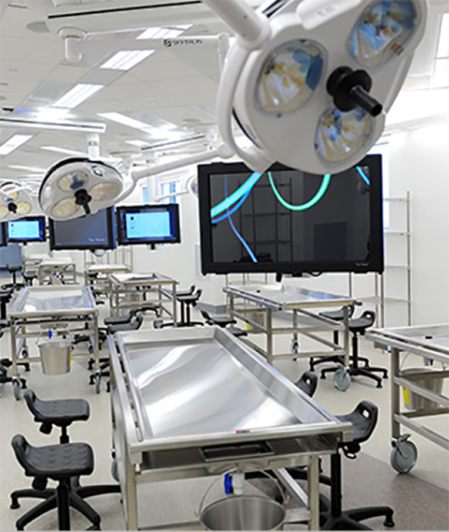
    Correlazione anatomica • Analisi cadaverica umana

    View Study

Continua a leggere

Biorigenerazione nella Miomodulazione

Bioregenerative

Myomodulation® provides a revolutionary approach to treat the aging appearance and organs ( skin, muscles) that will provide patients with more long-lasting and youthful results.

       Services Provided

  • Rejuvenation

  • Embellishment

  • Tissue Reshaping

To Resume

  • 3D Isovolumetric Tissular repositioning

  • Myoplasty

  • Myopexy

  • Myotension

  • Bioregeneration

The skin of the face is one of the areas most affected by the passage of time. It is even possible that it suffers from phenomena of premature aging if we do not take care of it properly. Crow's feet, sagging in the mouth and cheekbones, the bar-code effect, forehead wrinkles, all these effects and more are symptoms of aging of our skin. There are many ways to combat these phenomena, including MYOMODULATION techniques.

There is indeed a large amount of medico-aesthetic treatments to improve the appearance of the skin of the face and to find a younger appearance. There are also many treatments that can be associated with each other for an ever more effective result.

Continua a leggere

Istologia

Istologia

Su ratti Wistar

Per gentile concessione del Laboratorio di Patologia del Dipartimento di Medicina della Scuola di Ribeirão Preto – USP

istologia

01

Studio
Gruppo

Sperimentatori : R.Mené,M.Kim,A.Tenenbaum

N= 29 ratti Wistar
Peso medio : 421g

Origine degli animali : Biotério Central del Campus di Ribeirão Preto della Università di São Paulo
Manutenzione : Patologia Sperimentale Laboratorio del Dipartimento di Patologia della Scuola di Medicina di Ribeirão Preto – USP
Alloggio degli animali : gabbie in polipropilene
Alimentazione degli animali : dieta di base del laboratorio e acqua
Condizioni di luce : cicli naturali luce/buio in condizioni di base controllate
Sperimentatori : A.Tenenbaum,R.Mené,Kim

dipartimento di patologia

02

Sacrificio degli animali e campioni istologici

Microscopia convenzionale

I topi sono stati eutanizzati in camere a CO2.

  • Incisione della pelle con una lama n° 23
  • Individuazione e isolamento del muscolo pretibiale usando forbici Metzenbaum
  • Sezionamento dei tendini con forbici Mayo.
  • Fissazione di porzioni dei muscoli su sezioni di sughero
  • Inclusione utilizzando soluzione formolo al 50% - 10% tamponata e 50% di paraffina pura per lo studio microscopico ottico convenzionale.
  • Colorazione delle sezioni come processo istologico di routine.
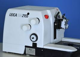
  • Microtome usato : Leika RM 2155
  • Colorazione delle sezioni muscolari con

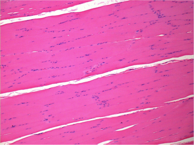
-ematossilina-eosina
-tricromia di Masson
-e picro-sirius rosso.

  • Esame istologico è stato effettuato con un microscopio convenzionale (Olympus BH-2).
  • Osservazione con lampada polarizzante
  • Documentazione fotografica è stata effettuata con un microscopio Leika DMR collegato a una fotocamera digitale Leika CD 300F e a un PC compatibile.
microtomo

03

Materiale

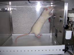
6 ratti/gabbia

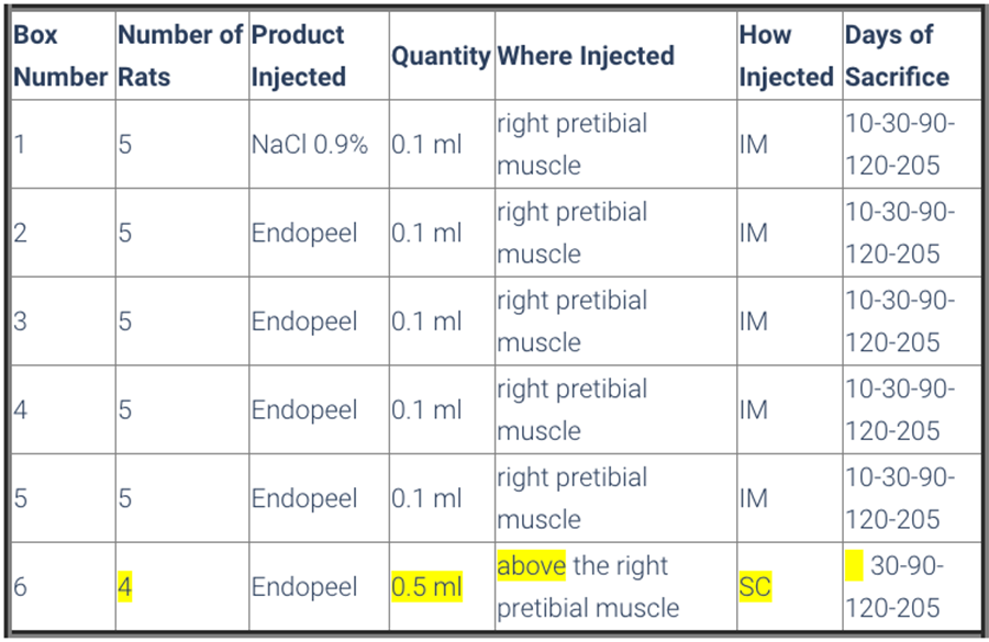
  • Gli animali sono stati divisi in 6 gabbie, in modo che le gabbie da 1 a 5 avevano 5 ratti e la gabbia numero 6 aveva 4 ratti 
  • Tutti gli animali sono stati sottoposti a una valutazione della deambulazione tramite impronte su carta speciale, con precedente immersione delle zampe posteriori in una soluzione di sapone. 
  • Gabbia 1 aveva il gruppo di controllo, che ha ricevuto l'applicazione di 0,1 ml di soluzione salina ( NaCl 0,9%) sul muscolo pretibiale destro. 
  • I topi delle gabbie 2 a 5 hanno ricevuto 0,1 ml di Endopeels Original Main Product sullo stesso muscolo del gruppo di controllo.

  • Gabbia 6 era costituita da 4 ratti, e ciascuno ha ricevuto 0,5 ml del prodotto principale Endopeels Original iniettato nel tessuto sottocutaneo. 

  • Tutti gli animali sono stati sottoposti a valutazione della deambulazione prima e dopo le rispettive iniezioni.

gabbie per ratti

04

Metodi

Gli animali vengono sacrificati dopo 10 giorni-1 mese-3 mesi-7 mesi

tabella del sacrificio

05

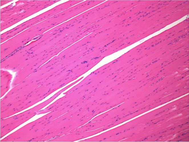
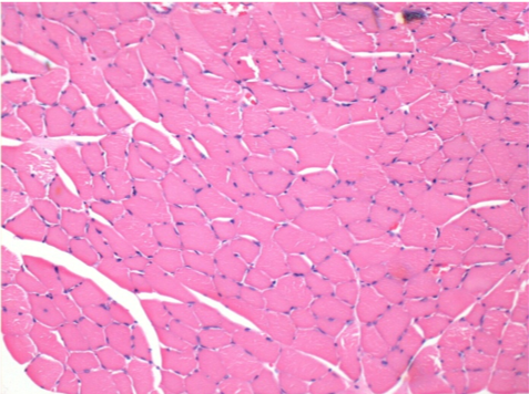
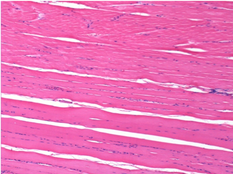
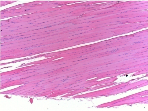
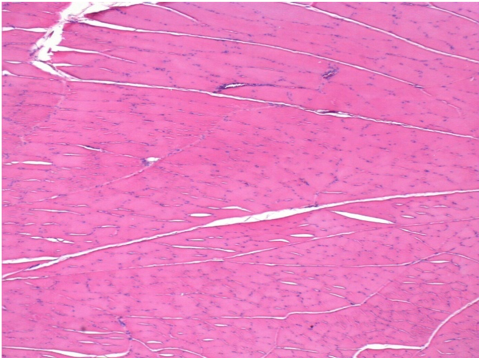
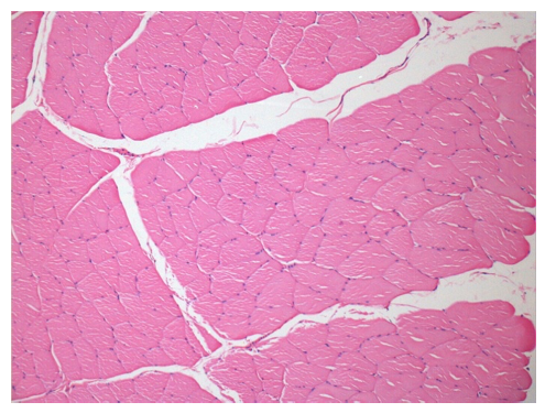
Controllo

Dopo l'iniezione di NaCl 0.5%

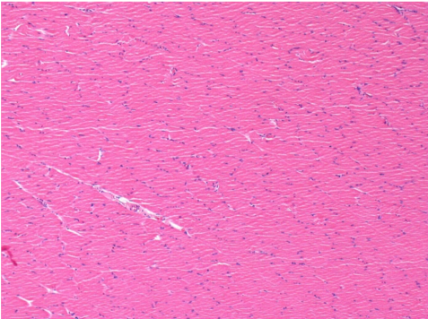
Commento : Nulla da segnalare dopo l'iniezione della soluzione salina

senza trattamento

L:Pretibiale-Nessun trattamento

R:Pretibiale-Dopo 0.1 ml NaCl 0.9% IM

06

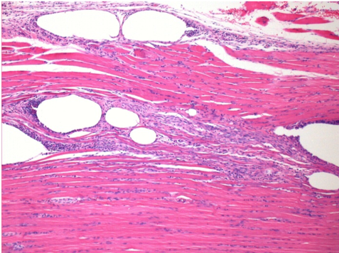
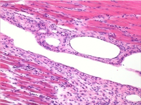
10 giorni dopo l'iniezione di Endopeel

Gabbia 2

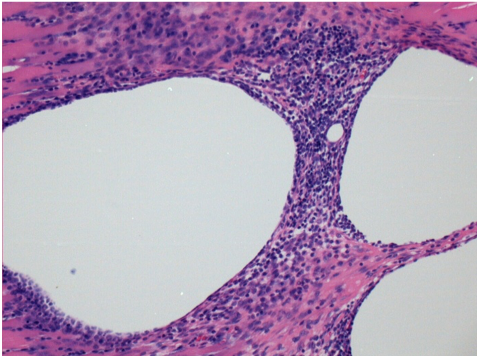
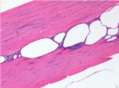
10 giorni dopo l'iniezione di Endopeel 0.1ml nel muscolo pretibiale destro.

Qui si può osservare la formazione delle vacuoli che sono circondate da linfociti. Le vacuoli sono diverse dalla necrosi tissutale . La presenza di linfociti è legata alla permeabilità delle membrane cellulari.


controllo sinistro- 100xD10

L : Controllo-100xD10

destro 100xD10

R:100xD10

destro200xx

R :200xD10

destro400xxD10

R :400xD10

07

1 mese dopo l'iniezione di Endopeel

Gabbia 3

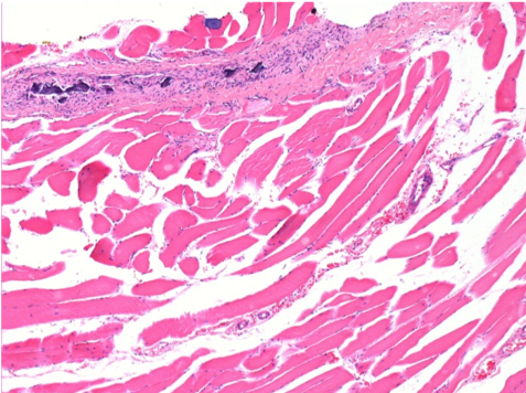
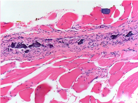
1 mese dopo l'iniezione di Endopeel 0.1ml nel muscolo pretibiale destro.
Ciò che si vede in nero nelle immagini non è una necrosi come potrebbero immaginare alcuni scienziati!
Infatti, devono essere considerate 4 conclusioni

  • un artefatto della colorazione
  • assenza di necrosi
  • apoptosi
  • un processo biorigenerativo 

Scopri di più sul processo bioregenerativo

controllo sinistro- 100xD30

L : Controllo-100xD30

destro 100x

R:100xD30

Definizione di necrosi

destro200xxD30

R :200xD30

destro400xxD30

R :400xD30

08

3 mesi dopo l'iniezione di Endopeel

Gabbia 4

3 mesi (D90) dopo l'iniezione di Endopeel 0.1ml nel muscolo pretibiale destro.

controllo sinistro- 100xD90

L : Controllo-100xD90

destro 100x

R:100xD90

09

7 mesi dopo l'iniezione di Endopeel

Gabbia 5

7 mesi (D210) dopo l'iniezione IM di Endopeel 0.1ml nel muscolo pretibiale destro.

Restitutio ad integrum completa dopo 7 mesi

controllo sinistro- 100xD210

L : Controllo-100xD210

destro 100x

R:100xD210

L Controllo 50xD210

L :Controllo 50xD210

R50X-D210

R50X-D210


10

Iniezione di Endopeel nel tessuto sottocutaneo

Gabbia 6

0.5 ml ( 5x 0.1ml)  Iniezione SC di Endopeel nella regione pretibiale sottocutanea destra.

L:200x-Controllo-SC

R-D10-SC-200X

R-D10-SC-200X

R-D30-SC-200X

R-D30-SC-200X

R-D90-SC-200X

R-D90-SC-200X

R-D210-SC-200X

R-D210-SC-200X

R-D210-SC-400X

R-D210-SC-400X


conclusioni
    • Endopeel induce una miofibrolisi selettiva e reversibile e una reazione infiammatoria per un periodo di circa 1 mese, approssimativamente
    • I cambiamenti muscolari sono reversibili nella quasi totalità
    • Il muscolo è il luogo migliore per iniettare Endopeel per maggiore efficacia, controllo e durata della sua azione
    • Non sono state riscontrate necrosi né ascessi durante lo studio.

Altro studio applicato alla Ginecologia

Leggi di più

Continua a leggere

Apoptosi

Apoptosis

Definition

  • in line with the body's metabolic and homeostasis plan

    for the defense and prevention of genetic mutations or viral attacks,which endanger the  survival of the organism

  • Apoptosis is a selective mechanism subjected to a regulatory plan of survival and death factors

    When DNA damage occurs,the factors are activated by the environment surrounding the damaged cell,as information from other nearby cells,which they receive from the damaged cell,the information needed to prepare the rescue plan.The signaling of survival factors reach the damaged cell,activating the genetic resident control factor program.In DNA transcriptional and regulatory factors attempt rescue,seeking to repair the damage.Programmed cell death factors are activated via caspases,which are proteins that form a scaffold at the level of the cytosolic plasma membrane,and they act by activating a proteolytic cascade regulated by phosphorylations.Mitochondrial cytochrome c,released into cytosol,participates in activation of caspases.

  • Apoptosis may be triggered by an external signal

    Phosphatidylserine moves from the position inherent in the cytosolic plasma membrane,going outside on the extracellular plasma membrane,and it is formed as if it were a receptor with external signaling factor on the cell in view of the macrophages,which intercept the signal positioning themselves on the cell,then incorporate it and degrade it in their body.


Main causes leading to programmed cell death

  • Oxidative stress

    caused by signaling factors that come from the external environment(magnetic radiation,reactive chemical compounds and gases).

  • Reactive factors

    such as Ros(highly reactive products of the partial reduction of O2,superoxide *O-2,minor byproduct of oxidative phosphorylation)

  • Viral infections or stress

    such as heat shock


Bioenergetic signaling mechanism of apoptosis

  • Signaling is information in the form of energy in living beings

    by animating metabolic cycles and therefore matter.

    Through chemical-biological evolution,the cell has managed to develop a constitutive plan,animated by complex energy processes,where matter is transformed through energy by regulating and making the metabolic systems of living beings function.Matter to be animated by energy,there must be an internal order in living beings,which needs an energy supply to make it work. This is a thermodynamic plane,which must ensure sufficient availability of energy,which is supplied and then extracted from the matter that animates the surrounding Cosmos.


  • Maintaining a certain type of order in the living organism

    that keeps it away from a equilibrium that can bring it to a steady state,responsible for the decay of the matter.


Apoptosis is made necessary

  • when damage in DNA jeopardizes a certain type of order

    consisting of a stationary-dynamic plane,which keeps away from the dynamic order of reactions

  • The road to old age is littered

    with a constant accumulation of mutations in the DNA,which the subsequent statistical increase in disease and cell senescence,directly proportional to the errors and damages that accumulate in the DNA or inside the macromolecules,regulated by biosynthesis.


Apoptosis & inflammation

  • Apoptosis is an effective means of preventing irreversible mutations

    that add up and are transmitted to the subsequent progeny of cells

  • Arachidonic acid is an essential component of Endopeel

    and this is manipulated in such a way that the entire molecular composition amplifies a cascade of reactions,which reactions initiate a renewal of the tissue that leads to an apoptotic process

  • Endopeel gives the necessary impetus to an energetic process

    that makes the mechanism of survival and death factors more selective.The selective process,on the one hand with survival factors,triggers an attempt to rescue the cell from mutations that could endanger the organism.If this rescue attempt does not work within a set time,phosphatidylserine from the inside of the plasma membrane slides outwards,capturing the signals triggering a caspase cascade ends the life of the cell,which is directed towards apoptosis.


  • Conclusion

    Apoptosis is an essential concept for slowing down the senescence process.

    As it is well described in the article "Journal of Advanced Research".The homeostasis processes between anti-inflammatory and inflammatory factors are directly linked to lifestyle,and therefore aging itself is also an accumulation of mutations.The disease that makes its way first is aging,the general structure suffers irreversible damage.The problem is precisely to arrive in time before these damages are irreversible.For this reason,apoptosis is an essential concept for slowing down the senescence process.

Ageing: Is there a role for arachidonic acid and other bioactive lipids ?

Do you still have questions left?

Questo indirizzo email è protetto dagli spambots. È necessario abilitare JavaScript per vederlo.

Continua a leggere