MECHANISTIC SCIENCE

MECHANISMS OF ACTION

FROM CHEMICAL INPUT TO
THREE-DIMENSIONAL MECHANICAL RESPONSE

Myomodulation converts a transient chemical input into a controlled mechanical response through the interaction of directional forces within an anisotropic and viscoelastic muscular system.

The muscle acts as a transfer function: chemical signaling is translated into vector–tensor coupling, three-dimensional tissue redistribution and the functional outputs of myoplasty, myopexy and myotension.

CHEMICAL INPUT Transient signal
→
TRANSFER FUNCTION Muscular response
→
3D OUTPUT Mechanical redistribution
MYOPLASTY 3D reshaping
MYOPEXY Spatial repositioning
MYOTENSION Tension modulation
MECHANISTIC PATHWAY
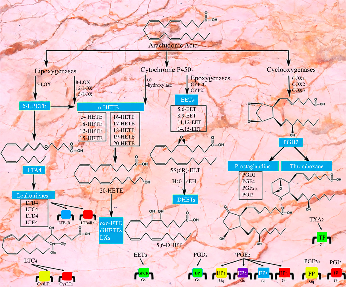
Scientific diagram illustrating biochemical signaling pathways associated with Myomodulation mechanisms of action
CHEMICAL SIGNAL → MUSCLE TRANSFER FUNCTION → MECHANICAL RESPONSE
CORE PRINCIPLE Transient molecular interaction can generate a persistent mechanical response without a permanent volumizing depot.
SCIENTIFIC NAVIGATION

SCIENTIFIC CONTENTS

Navigate through the complete mechanistic framework of Myomodulation, from chemical input and tissue interaction to biomechanical response, experimental evidence and clinical expression.

MECHANISTIC ROADMAP
CHEMICAL INPUT → MUSCLE TRANSFER FUNCTION → V⊥ / T∥ → ANISOTROPIC + VISCOELASTIC RESPONSE → 3D REDISTRIBUTION → MYOPLASTY · MYOPEXY · MYOTENSION
MECHANISTIC OVERVIEW

FROM CHEMICAL INPUT TO MECHANICAL OUTPUT

Myomodulation begins with a transient chemical input and culminates in a controlled mechanical response. The injected formulation does not act as a volumizing filler or permanent implant. Instead, it initiates local tissue interactions that are translated by the muscular system into directional changes in tone, geometry and three-dimensional tissue organization.

01
CH
CHEMICAL INPUT

Transient Local Signal

A stereochemically controlled formulation is delivered in the subcutaneous plane, producing localized physicochemical interaction without creating a permanent volumizing depot.

LOCAL TRANSIENT NON-VOLUMIZING
→
02
TF
MUSCLE TRANSFER FUNCTION

Biological Translation

Muscle architecture, fiber orientation, resting tone, anisotropy and viscoelastic behavior determine how the transient chemical input is converted into a spatially organized mechanical response.

ARCHITECTURE DIRECTION TONE
→
03
3D
MECHANICAL OUTPUT

Controlled 3D Response

Directional mechanical outputs redistribute projection, tissue tension and local geometry through vector–tensor coupling without requiring persistent injected material.

RESHAPING REPOSITIONING TENSION
MECHANISTIC LOGIC
CHEMICAL SIGNAL
→
MUSCLE TRANSFER FUNCTION
→
DIRECTIONAL RESPONSE
→
3D REDISTRIBUTION
MYOPLASTY

Three-dimensional reshaping of muscle-related geometry and projection.

MYOPEXY

Controlled spatial repositioning and translation of tissue projection.

MYOTENSION

Modulation of resting tension while functional contractile activity is preserved.

CORE CONCEPT
The chemical input is transient. The mechanical response is architecture-dependent and may persist after molecular clearance.
BIOLOGICAL TRANSLATION

MUSCLE AS A TRANSFER FUNCTION

The muscular system does not behave as a passive target. It transforms a transient chemical input into a directionally organized mechanical response. The resulting output depends on local anatomy, fiber orientation, resting tone, anisotropy and viscoelastic behavior.

INPUT
CH

Transient Chemical Signal

Local physicochemical interaction initiated after controlled subcutaneous administration.

LOCAL
TRANSIENT
NON-VOLUMIZING
→
TRANSFER FUNCTION
f(tissue)

MUSCLE

An active, anisotropic and viscoelastic biological system that converts chemical signaling into mechanical behavior.

OUTPUT = f [ architecture · orientation · tone · anisotropy · viscoelasticity ]
→
OUTPUT
3D

Mechanical Response

Directionally organized deformation, redistribution of projection and modulation of tissue tension.

DIRECTIONAL
MECHANICAL
3D
01

MUSCLE ARCHITECTURE

Local geometry determines how mechanical loading is distributed through the treated muscular region.

02

FIBER ORIENTATION

Directional organization of muscle fibers contributes to an anisotropic response rather than uniform deformation.

03

RESTING TONE

Baseline muscular tension influences the mechanical state from which myotension and reshaping develop.

04

ANISOTROPY

Tissue response varies with direction. The same mechanical input does not generate identical deformation along every axis.

05

VISCOELASTICITY

The muscular response combines immediate deformation with a time-dependent biological and mechanical adaptation.

KEY PRINCIPLE
A given chemical input does not prescribe a fixed shape. The final mechanical output is determined by the transfer properties of the treated muscular system.
2-MUSCLE AS A TRANSFER FUNCTION
BIOPHYSICAL FRAMEWORK

VECTOR NORMAL-TO-PLANE ACTION

In the Myomodulation model, the vector defines a single directional axis perpendicular to the local muscular plane. It establishes the normal deformation direction and must not be confused with tangential or in-plane tissue displacement.

01
GEOMETRIC DEFINITION

One Direction

The vector is unidirectional. It defines a local axis of mechanical action rather than a multidirectional field.

V⊥
LOCAL MUSCULAR PLANE
V⊥
MUSCULAR PLANE
NORMAL DIRECTION
V⊥ normal to the local muscular plane
02
MECHANICAL ROLE

Deformation Axis

The vector establishes the local orthogonal axis along which projection and normal deformation can be expressed.

NORMAL AXIS
01

PERPENDICULAR TO THE PLANE

The vector is oriented normal to the local muscular surface. Its defining property is orthogonality.

02

DEFINES LOCAL DEFORMATION

It establishes the directional axis on which local three-dimensional deformation is organized.

03

NO IN-PLANE TRANSLATION

The vector does not describe tangential transfer across the muscular plane. That in-plane organization belongs to the tensor field.

VECTOR
V⊥
Normal direction
≠
TENSOR
T∥
In-plane directional organization
KEY PRINCIPLE
Vector = normal direction. It defines the local deformation axis; it does not describe in-plane tissue transfer.
BIOPHYSICAL FRAMEWORK

TENSOR IN-PLANE DIRECTIONAL FIELD

In the Myomodulation model, the tensor organizes directional action within the local muscular plane. Unlike the vector, it does not define a single perpendicular axis. It coordinates in-plane tissue transfer and contributes to the spatial redistribution of shape, tension and projection.

01
GEOMETRIC DEFINITION

In-Plane Organization

The tensor acts within the local muscular plane. Its role is to organize directional relationships between mapped points rather than define one single orthogonal axis.

T∥
DIRECTIONAL FIELD WITHIN THE MUSCULAR PLANE
LOCAL MUSCULAR PLANE
T∥
T∥ directional organization within the local muscular plane
02
MECHANICAL ROLE

Tissue Redistribution

The tensor coordinates tangential tissue displacement between mapped points and organizes the in-plane component of the three-dimensional mechanical response.

DIRECTIONAL FIELD
01

OPERATES WITHIN THE PLANE

Tensor action is tangential to the local muscular surface and remains organized within that plane.

02

MULTIDIRECTIONAL ORGANIZATION

It represents a field of directional relationships rather than a single one-way action.

03

TRANSFERS TISSUE BETWEEN POINTS

Its functional role is to organize tangential redistribution across the mapped tissue grid.

STARTING POINT
Mapped Grid Defined anatomical coordinates
→
TENSOR FIELD
T∥ In-plane directional organization
→
RESULT
Redistribution Controlled tangential tissue transfer
VECTOR
V⊥
One normal direction
≠
TENSOR
T∥
In-plane directional field
KEY PRINCIPLE
Tensor = in-plane directional organization. It coordinates tangential tissue redistribution and prepares the local geometry for coupled three-dimensional deformation.
MATERIAL RESPONSE

ANISOTROPIC & VISCOELASTIC GRID RESPONSE

Skeletal muscle behaves as an active, anisotropic and viscoelastic biological material. Within the treatment grid, each local cell represents a deformable tissue unit whose mechanical response depends on direction, local architecture, resting tone and time-dependent tissue behavior.

01
BASELINE

Resting Grid Cell

Before loading, the local tissue unit has its own geometry, fiber orientation, resting tone and baseline mechanical state.

RESTING STATE LOCAL ARCHITECTURE
DIRECTIONAL
LOADING
→
02
COUPLED INPUT

Local Mechanical Loading

V⊥
T∥

Normal and in-plane directional components act on a biological material whose deformation is neither uniform nor instantaneous.

V⊥ T∥ LOCAL STRAIN
3D
STRAIN
→
03
RESPONSE

Deformed Grid Cell

The local cell changes shape according to directional loading and material properties, producing a three-dimensional strain response without requiring added material volume.

SHAPE CHANGE VOLUME PRESERVED
A
DIRECTION-DEPENDENT RESPONSE

ANISOTROPY

Muscle does not respond identically along every axis. Fiber orientation, fascial organization and local architecture modify how an applied load is transmitted and how each grid cell deforms.

RESPONSE ≠ SAME IN ALL DIRECTIONS
VE
TIME-DEPENDENT RESPONSE

VISCOELASTICITY

The mechanical response combines an immediate deformational component with time-dependent adaptation. Tissue behavior therefore depends not only on load magnitude and direction, but also on time.

RESPONSE = DIRECTION + TIME
CONCEPTUAL CONSTITUTIVE RELATIONSHIP
ε3D(x, y, z, t) = f [ V⊥, T∥, tissue anisotropy, resting tone ]
Conceptual mechanistic relationship — not a patient-specific quantitative material model.
MECHANICAL OUTPUT
MYOPLASTY
MYOPEXY
MYOTENSION
KEY PRINCIPLE
The grid does not deform uniformly. Each local tissue unit responds according to directional loading, tissue anisotropy, resting tone and viscoelastic behavior.
COUPLED MECHANICS

VECTOR–TENSOR COUPLING & 3D REDISTRIBUTION

Three-dimensional reshaping emerges from the coupling of two distinct geometric components: a vector acting normal to the muscular plane and an in-plane directional tensor field. Their interaction organizes local deformation and tissue redistribution within an anisotropic and viscoelastic biological system.

NORMAL COMPONENT
V⊥

VECTOR

Defines the local deformation axis normal to the muscular plane.

ONE NORMAL DIRECTION
COUPLES WITH
×
IN-PLANE COMPONENT
T∥

TENSOR

Organizes directional relationships and tangential tissue transfer within the muscular plane.

DIRECTIONAL FIELD
CONCEPTUAL COUPLING
V⊥ normal vector
→
T(V)
→
R⃗ vectorial mechanical response
Conceptual tensor–vector coupling. It is not a scalar addition and not an ordinary cross product.
THREE-DIMENSIONAL RESPONSE

Redistribution Rather Than Added Volume

The coupled response modifies local geometry by redistributing existing tissue volume between spatial dimensions. Length, width and projection may increase, decrease or remain relatively stable according to the intended three-dimensional architecture, while overall material volume is preserved.

L, W and H are target-dependent variables; their reciprocal redistribution preserves the overall tissue volume.
L ↕ may decrease, increase or remain stable
W ↕ may increase, decrease or remain stable
H ↕ projection is redistributed according to the target geometry
V = preserved
COUPLED 3D DEFORMATION
V⊥
LOCAL GRID CELL
01

MYOPLASTY

Three-dimensional modification of muscular geometry and projection.

02

MYOPEXY

Spatial translation and controlled repositioning of tissue projection.

03

MYOTENSION

Modulation of resting muscular tension while contractile activity is preserved.

MECHANICAL SYNTHESIS
V⊥ ∗ T∥ → ANISOTROPIC / VISCOELASTIC RESPONSE → 3D REDISTRIBUTION
∗ denotes conceptual vector–tensor coupling here; it does not imply a formally defined mathematical convolution operator.
KEY PRINCIPLE
V⊥ defines the normal axis; T∥ organizes the in-plane field. Their coupling produces a directional three-dimensional mechanical response that redistributes existing tissue geometry without requiring added volume.
BIOLOGICAL EVIDENCE

EXPERIMENTAL & HISTOLOGIC SUPPORT

Histologic and experimental observations provide biological support for a transient local tissue response following Myomodulation. Human tolerance observations and preclinical histology must, however, be interpreted separately and within the limits of their respective experimental models.

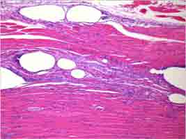
PRECLINICAL HISTOLOGY
Preclinical histologic section showing localized vacuolization in treated tissue
MICROSTRUCTURAL RESPONSE
Preclinical histology. Localized vacuolization observed after exposure, followed by structural restitution in subsequent observations. The finding illustrates a transient microstructural response and should be interpreted within the limits of the experimental model.
01
HUMAN OBSERVATIONS

Early Tissue Tolerance

Early human histologic observations support preserved tissue architecture and local tolerance in the examined material.

NO COAGULATIVE NECROSIS
NO THROMBOSIS
NO HEMORRHAGE
These observations describe the examined histologic material and should not be generalized beyond the available evidence.
02
PRECLINICAL OBSERVATIONS

Localized Vacuolization & Restitution

Experimental tissue sections have shown localized vacuolization after exposure, followed in subsequent observations by structural restitution.

1 EXPOSURE
→
2 VACUOLIZATION
→
3 RESTITUTION
The sequence is compatible with a transient microstructural response, but does not by itself establish the complete mechanism responsible for the clinical effect.
EVIDENCE INTERPRETATION

What Histology Contributes to the Mechanistic Model

01

LOCALIZED RESPONSE

Histologic findings are compatible with a spatially localized tissue interaction rather than a persistent volumizing depot.

02

TRANSIENT MICROSTRUCTURAL CHANGE

Vacuolization followed by restitution supports the concept of a reversible or transient microstructural event in the experimental tissue model.

03

PRESERVED TISSUE INTEGRITY

The available human observations are consistent with tissue tolerance and preserved structural integrity in the examined samples.

THE EVIDENCE SUPPORTS
  • A localized biological tissue response
  • Transient microstructural modification in preclinical material
  • Subsequent structural restitution in the observed experimental sequence
  • Early histologic tissue tolerance in the examined human material
THE EVIDENCE DOES NOT BY ITSELF PROVE
  • A complete molecular mechanism of action
  • That vacuolization alone explains the clinical effect
  • A direct quantitative relationship between histologic change and three-dimensional clinical reshaping
  • That preclinical findings can be transferred without qualification to every human tissue or indication
MECHANISTIC BRIDGE
LOCAL TISSUE INTERACTION → TRANSIENT MICROSTRUCTURAL RESPONSE → RESTORED TISSUE INTEGRITY → FUNCTIONAL MECHANICAL SUPPORT
This sequence represents a conceptual interpretation of the available observations, not a fully validated causal pathway.
KEY PRINCIPLE
Histology supports a transient local tissue response and subsequent restitution. It contributes biological evidence to the mechanistic framework without, by itself, proving the complete pathway responsible for the lasting clinical mechanical effect.
MOLECULAR KINETICS VS CLINICAL RESPONSE

CLEARANCE & DURATION OF ACTION

Molecular persistence and clinical duration are not equivalent. The components involved in the initial physicochemical interaction are cleared on substantially shorter timescales than the mechanical response observed clinically.

MOLECULAR PRESENCE
tmolecular

Hours to weeks

≠
DIFFERENT TIMESCALES
CLINICAL RESPONSE
tclinical

May persist for months

01
INITIAL PHASE
t₀

Local Physicochemical Input

Controlled subcutaneous administration initiates the local physicochemical interaction that precedes the mechanical response.

TRANSIENT INPUT
02
AROMATIC ACID
~24 h

Rapid Clearance

According to the available formulation data, the aromatic acid component undergoes near-complete elimination within approximately twenty-four hours.

SHORT MOLECULAR PRESENCE
03
LIPID CARRIER
~3 weeks

Longer Carrier Kinetics

The arachidonic-acid-containing lipid carrier follows a longer clearance timescale, extending to approximately three weeks.

TRANSIENT CARRIER PHASE
04
CLINICAL RESPONSE
MONTHS

Persistent Mechanical Effect

Clinical reshaping, repositioning and tension modulation may remain observable for months, despite clearance of the initially administered molecular components.

RESPONSE > MOLECULAR PRESENCE
TEMPORAL DECOUPLING

Clearance Does Not Define Clinical Duration

The persistence of the clinical effect should therefore not be interpreted as persistence of the injected preparation within the tissue.

AROMATIC ACID
~24 h
LIPID CARRIER
~3 weeks
CLINICAL EFFECT
months
HOURS DAYS WEEKS MONTHS
MECHANISTIC INTERPRETATION

Why Can the Mechanical Response Outlast Molecular Presence?

01

TRANSIENT INPUT

The administered preparation provides an initial local physicochemical input rather than a permanent structural implant.

02

BIOLOGICAL TRANSFER

The muscular system converts that input through its architecture, resting tone, anisotropy and viscoelastic properties.

03

MECHANICAL REORGANIZATION

The resulting response involves spatial redistribution and modification of tissue geometry rather than persistence of injected material.

04

CLINICAL PERSISTENCE

The mechanically reorganized state may remain clinically observable after the initial molecular components have been cleared.

TEMPORAL MODEL
CHEMICAL INPUT → TRANSIENT TISSUE INTERACTION → MECHANICAL RESPONSE → MOLECULAR CLEARANCE ≠ END OF CLINICAL EFFECT
CLINICAL VARIABILITY

Duration Is Not a Fixed Biological Constant

The persistence and magnitude of the clinical response can vary according to the treated anatomy, local muscular architecture, baseline tissue characteristics, functional loading and treatment strategy.

ANATOMY MUSCLE ARCHITECTURE RESTING TONE FUNCTIONAL LOADING TREATMENT GEOMETRY
KEY PRINCIPLE
Molecular presence ≠ clinical duration. The aromatic acid is cleared rapidly and the lipid carrier over a longer but still limited period, while the mechanically expressed clinical response may persist for months.
FUNCTIONAL CLINICAL EXPRESSION

CLINICAL MECHANICAL OUTPUTS

The coupled mechanical response can be expressed clinically through three complementary outputs: three-dimensional reshaping, spatial repositioning and modulation of resting muscular tension. Their relative contribution depends on anatomy, treatment geometry and the intended aesthetic or functional objective.

01
3D
GEOMETRIC OUTPUT

MYOPLASTY

THREE-DIMENSIONAL RESHAPING

Myoplasty describes controlled modification of muscle-related three-dimensional geometry. Existing tissue volume is redistributed to modify contour, projection and local shape without requiring volumetric augmentation.

RESHAPING PROJECTION CONTOUR
02
↗
SPATIAL OUTPUT

MYOPEXY

SPATIAL REPOSITIONING

Myopexy describes directional repositioning of muscle-related projection and tissue geometry. The point of maximal projection can be translated within the treated architecture according to the desired three-dimensional result.

TRANSLATION REPOSITIONING DIRECTION
03
τ
TENSION OUTPUT

MYOTENSION

RESTING TENSION MODULATION

Myotension describes modulation of resting muscular tension while functional contractile activity is preserved. The intended effect is not paralysis, but modification of the baseline mechanical state.

RESTING TONE FUNCTION PRESERVED NO PARALYSIS
CLINICAL SYNTHESIS
MYOPLASTY ↔ MYOPEXY ↔ MYOTENSION

These outputs are not mutually exclusive. A single treatment strategy may combine reshaping, repositioning and tension modulation in different proportions according to the local anatomy and target geometry.

ISOVOLUMETRIC GEOMETRY

Shape Changes Without Added Volume

Myomodulation is based on redistribution of existing tissue geometry. Length, width and projection can each increase, decrease or remain relatively stable depending on the desired morphology, while overall material volume remains preserved.

V ≈ L × W × H = constant
L
↕
LENGTH

May decrease, increase or remain relatively stable.

W
↕
WIDTH

May increase, decrease or remain unchanged according to the target contour.

H
↕
PROJECTION

Redistribution determines the location and magnitude of projection.

V
=
VOLUME

Preserved within the isovolumetric reshaping model.

TARGET-DEPENDENT RESPONSE

The Desired Geometry Determines the Output

There is no single predetermined shape. The directional strategy is adapted to the morphology to be preserved, reduced, expanded, translated or redistributed.

01

WIDTH PRESERVATION

Width may remain relatively stable when the objective is to preserve the existing lateral architecture.

02

WIDTH INCREASE

Width may be increased when lateral expansion or reduction of an external concavity is part of the intended morphology.

03

WIDTH REDUCTION

Width may also be reduced when a narrower architecture is the intended mechanical and aesthetic target.

FUNCTIONAL DISTINCTION

Mechanical Modulation Without Paralysis

↑ RESTING TONE
+
✓ CONTRACTILE ACTIVITY PRESERVED
≠
PARALYTIC NEUROMODULATION
CLINICAL EXPRESSION OF THE MECHANISTIC MODEL
VECTOR–TENSOR COUPLING → ANISOTROPIC / VISCOELASTIC RESPONSE → 3D REDISTRIBUTION → MYOPLASTY · MYOPEXY · MYOTENSION
KEY PRINCIPLE
The clinical output is target-dependent rather than shape-prescriptive. Myoplasty, myopexy and myotension combine to redistribute existing tissue geometry and mechanical tension according to the intended three-dimensional architecture.
MECHANISTIC SYNTHESIS

INTEGRATED MECHANISTIC MODEL

Myomodulation can be represented as a multistage mechanistic framework in which a transient chemical input is translated by active muscular tissue into a directionally organized mechanical response. The final clinical expression emerges from tissue-specific vector–tensor coupling, anisotropic and viscoelastic behavior, and three-dimensional redistribution of existing tissue geometry.

FROM INPUT TO CLINICAL OUTPUT
01
CH
TRANSIENT INPUT

CHEMICAL INPUT

Controlled subcutaneous administration initiates a localized physicochemical interaction. The injected preparation acts as a transient input and is not intended to function as a permanent volumizing depot.

LOCAL TRANSIENT NON-VOLUMIZING
↓ BIOLOGICAL TRANSLATION
02
f
ACTIVE BIOLOGICAL SYSTEM

MUSCLE AS A TRANSFER FUNCTION

Skeletal muscle behaves as an active, anisotropic and viscoelastic biological material. Local architecture, fiber orientation, resting tone and time-dependent material behavior determine how the transient input is translated into mechanical response.

ARCHITECTURE ANISOTROPY RESTING TONE VISCOELASTICITY
↓ DIRECTIONAL ORGANIZATION
03
GEOMETRIC ORGANIZATION

VECTOR–TENSOR COUPLING

NORMAL COMPONENT
V⊥

VECTOR

Defines the conceptual deformation axis normal to the local muscular plane.

CONCEPTUAL COUPLING
T(V)
→
R⃗

vectorial mechanical response

IN-PLANE COMPONENT
T∥

TENSOR

Organizes directional relationships and redistribution within the local muscular plane.

T(V) represents a conceptual tensor–vector coupling. It is not a scalar addition and not an ordinary cross product.
↓ MATERIAL RESPONSE
04
ε
ACTIVE MATERIAL RESPONSE

ANISOTROPIC & VISCOELASTIC DEFORMATION

Local tissue units do not deform uniformly. Directional loading is filtered by muscle architecture, anisotropy, baseline tension and viscoelastic behavior, creating a spatially and temporally organized three-dimensional response.

DIRECTION-DEPENDENT TIME-DEPENDENT 3D RESPONSE
CONCEPTUAL CONSTITUTIVE RELATION
ε3D(x,y,z,t) = f [ V⊥, T∥, tissue anisotropy, resting tone ]

Conceptual mechanistic representation — not a patient-specific quantitative material model.

↓ GEOMETRIC REDISTRIBUTION
05
ISOVOLUMETRIC RESPONSE

THREE-DIMENSIONAL REDISTRIBUTION

The resulting mechanical response redistributes existing tissue geometry rather than adding volume. Length, width and projection are target-dependent variables and may increase, decrease or remain relatively stable according to the intended architecture.

V ≈ L × W × H = constant
L ↕ LENGTH
W ↕ WIDTH
H ↕ PROJECTION
V = PRESERVED
↓ CLINICAL EXPRESSION
06
CLINICAL MECHANICAL OUTPUT

THREE COMPLEMENTARY EXPRESSIONS

3D

MYOPLASTY

3D RESHAPING

Modification of muscle-related geometry, contour and projection.

↗

MYOPEXY

SPATIAL REPOSITIONING

Directional translation of tissue projection within the treated architecture.

τ

MYOTENSION

TENSION MODULATION

Modulation of resting muscular tension while contractile activity remains preserved.

COMPLETE MECHANISTIC SEQUENCE
CHEMICAL INPUT → MUSCLE TRANSFER FUNCTION → V⊥ / T∥ COUPLING → ANISOTROPIC + VISCOELASTIC RESPONSE → 3D REDISTRIBUTION → MYOPLASTY · MYOPEXY · MYOTENSION
TRANSIENT INPUT
MOLECULAR CLEARANCE

The molecular components are eliminated on timescales considerably shorter than the potential duration of the clinical response.

≠
PERSISTENT OUTPUT
CLINICAL DURATION

Persistence of the mechanical result does not require persistence of the injected material within the tissue.

SCIENTIFIC INTERPRETATION
This is an integrated conceptual mechanistic model. Experimental histology, physiology and clinical observations provide support for individual elements of the framework, but the complete vector–tensor pathway should not be interpreted as a fully validated quantitative constitutive model.
KEY PRINCIPLE
Transient chemical input → architecture-dependent mechanical output. Myomodulation describes a directional, isovolumetric redistribution of tissue geometry in which muscle functions as the biological transfer system linking local physicochemical interaction to myoplasty, myopexy and myotension.
NEUROMUSCULAR EVIDENCE

NEUROMUSCULAR & ELECTROPHYSIOLOGIC EVIDENCE

Experimental observations at the neuromuscular and electrophysiologic levels provide additional support for the functional interpretation of Myomodulation. These findings should be considered as complementary evidence rather than as isolated proof of the complete mechanistic model.

01
EXPERIMENTAL MICROSCOPY

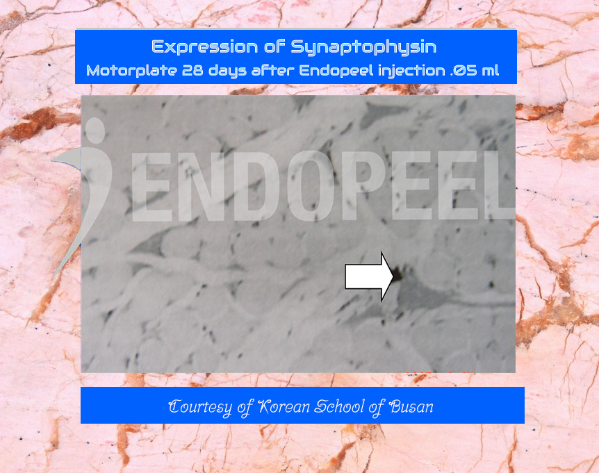
SYNAPTOPHYSIN EXPRESSION

Experimental image illustrating Synaptophysin expression after treatment
MOTOR-PLATE OBSERVATION
Historical experimental material illustrating Synaptophysin expression in relation to the motor-plate environment after treatment. The image should be interpreted as supportive experimental observation rather than as a standalone demonstration of the full mechanism of action.
MECHANISTIC INTERPRETATION

Synaptophysin is associated with presynaptic vesicular structures. Changes observed in experimental material may indicate a local neuromuscular response within the treated environment, but they should not be interpreted in isolation as evidence of neuromuscular blockade or paralysis.

02
EXPERIMENTAL ELECTROPHYSIOLOGY

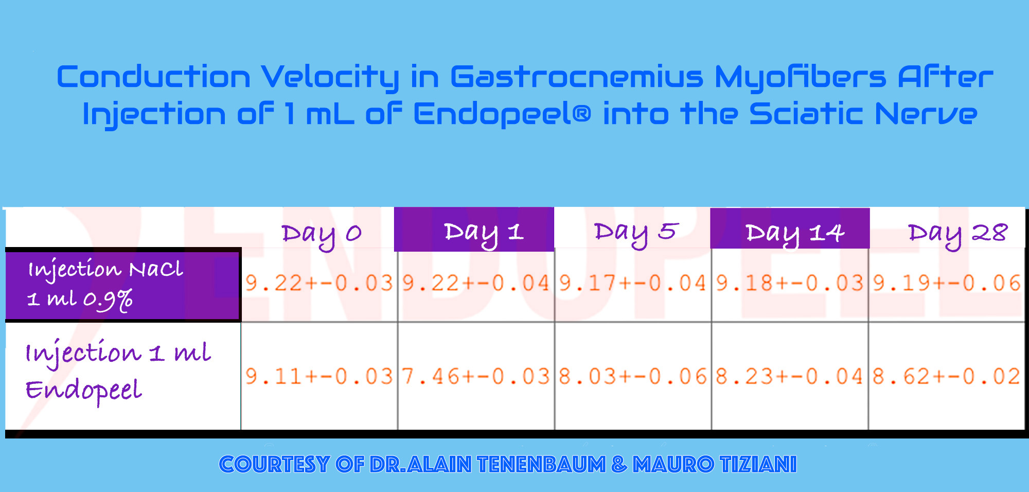
CONDUCTION VELOCITY

Experimental conduction velocity data in gastrocnemius muscle fibers after treatment
GASTROCNEMIUS EXPERIMENT
Experimental conduction-velocity observations in gastrocnemius muscle fibers over time following treatment. The data are presented as historical electrophysiologic evidence and should be interpreted within the limits of the experimental protocol.
FUNCTIONAL INTERPRETATION

The electrophysiologic observations are relevant because the clinical concept of Myomodulation is not based on suppression of muscle activity. Functional contractility is intended to remain preserved while resting mechanical behavior is modulated.

FUNCTIONAL DISTINCTION

Modulation of Resting Tone Without Paralysis

The experimental framework is consistent with a distinction between modification of resting muscular behavior and pharmacologic neuromuscular blockade.

↑
RESTING TONE

Experimental physiology supports an increase or modulation of the resting mechanical state of the treated muscle.

✓
CONTRACTILE FUNCTION

Muscle activity remains functionally preserved rather than being intentionally suppressed.

≠
NEUROMUSCULAR BLOCKADE

The intended effect differs fundamentally from paralytic neuromodulation.

EVIDENCE LOGIC
NEUROMUSCULAR OBSERVATION + ELECTROPHYSIOLOGIC OBSERVATION → PRESERVED FUNCTION + MODULATED RESTING STATE
The “+” symbols indicate complementary evidence domains, not a mathematical operation.
THESE OBSERVATIONS SUPPORT
  • The concept of a functional neuromuscular response
  • Preserved contractile activity rather than intended paralysis
  • A distinction between resting-tone modulation and neuromuscular blockade
  • A broader biological basis for the mechanical output model
THEY DO NOT BY THEMSELVES PROVE
  • The complete molecular pathway of Myomodulation
  • A direct causal relationship between Synaptophysin expression and clinical reshaping
  • A universal electrophysiologic response in every muscle or indication
  • The complete vector–tensor model without additional evidence
CONNECTION TO THE MECHANICAL MODEL
TRANSIENT LOCAL INPUT → NEUROMUSCULAR RESPONSE → RESTING-TONE MODULATION → PRESERVED CONTRACTILITY → MECHANICAL EXPRESSION
KEY PRINCIPLE
Myomodulation is not based on muscular paralysis. The available neuromuscular and electrophysiologic observations are compatible with modulation of the resting mechanical state while functional contractile activity remains preserved.
FROM EVIDENCE TO VISIBLE OUTCOME

FROM EXPERIMENTAL OBSERVATION TO CLINICAL EXPRESSION

Experimental tissue observations and visible clinical outcomes belong to different levels of evidence. The former can inform mechanistic interpretation; the latter illustrate the clinical expression of the resulting tissue response.

TRANSLATIONAL FRAMEWORK
EXPERIMENTAL OBSERVATION → TISSUE / MECHANICAL INTERPRETATION → CLINICAL EXPRESSION
Clinical photography illustrates outcome; it does not independently establish causation or prove the underlying mechanistic pathway.
01
EXPERIMENTAL + CLINICAL CORRELATION

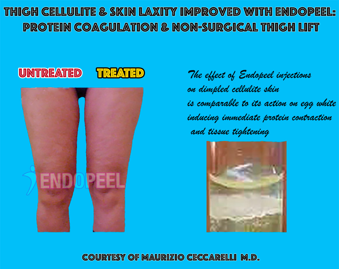
TISSUE RESPONSE & THIGH CONTOUR

Historical experimental and clinical material illustrating tissue and thigh contour observations
HISTORICAL EXPERIMENTAL MATERIAL
Historical material combining clinical contour observation with experimental tissue findings. It illustrates the attempt to relate local tissue-level modification to visible changes in thigh surface contour and laxity.
INTERPRETATION

The image is compatible with a model in which localized tissue interaction modifies the mechanical behavior of the treated region, with visible consequences for contour, firmness and surface architecture.

This association should be interpreted as supportive clinical correlation rather than proof that a single histologic process directly causes the visible outcome.
02
CLINICAL EXPRESSION

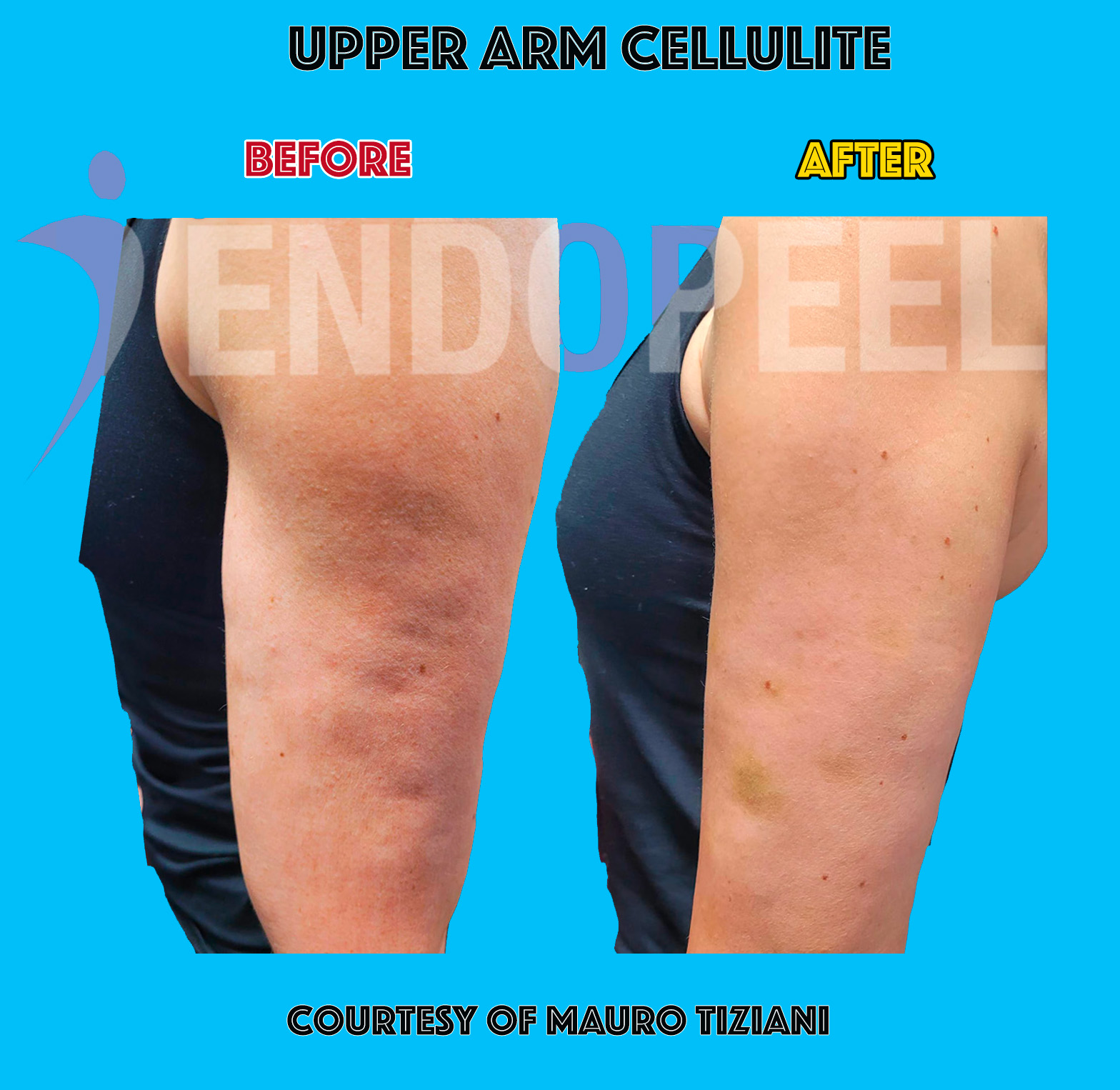
UPPER-ARM CONTOUR & FIRMNESS

Clinical before-and-after observation of upper-arm contour and skin laxity
CLINICAL OUTCOME
Clinical before-and-after material illustrating visible change in upper-arm contour and surface laxity after treatment.
MECHANICAL EXPRESSION

The visible result can be interpreted as the clinical expression of altered local tissue tension, support and three-dimensional contour rather than as simple volumetric filling.

CONTOUR FIRMNESS TENSION NON-VOLUMIZING
HOW THE LEVELS OF EVIDENCE CONNECT

Observation, Interpretation and Clinical Expression

01

OBSERVATION

Histologic, microscopic or physiologic findings describe what is observed within a specific experimental setting.

02

INTERPRETATION

These findings can support a mechanistic hypothesis involving changes in local tissue behavior, architecture and mechanical response.

03

CLINICAL EXPRESSION

Visible changes in contour, firmness, projection or tension represent the clinical phenotype of the resulting mechanical response.

SECONDARY CLINICAL OBSERVATION

PERIORBITAL APPEARANCE

Historical clinical material also includes observations in the periorbital region. Such images may illustrate visible changes in local appearance, but they should not be used to define the central muscular mechanism of Myomodulation.

This observation is retained as complementary clinical material, not as a primary mechanistic endpoint.
Historical clinical observation of periorbital appearance before and after treatment
Historical periorbital clinical observation.
MECHANICAL TRANSLATION
LOCAL TISSUE INTERACTION → MODIFIED TENSION / SUPPORT → 3D CONTOUR REDISTRIBUTION → VISIBLE CLINICAL CHANGE
CLINICAL IMAGES CAN SHOW
  • Change in visible contour
  • Change in apparent firmness or laxity
  • Redistribution of projection
  • Expression of a treatment-associated mechanical outcome
CLINICAL IMAGES CANNOT ALONE PROVE
  • The exact molecular pathway
  • A direct causal role for one histologic finding
  • The complete vector–tensor model
  • Generalization to every anatomy or indication
KEY PRINCIPLE
Experimental evidence and clinical photography answer different questions. Experimental observations help build the mechanistic framework; clinical images demonstrate its visible expression. Their interpretation is strongest when the distinction between evidence, hypothesis and outcome remains explicit.
SCIENTIFIC FAQ

FREQUENTLY ASKED QUESTIONS

Key scientific questions about Myomodulation, its mechanical framework, tissue response, duration of action and clinical interpretation.

01
What is the core mechanism proposed for Myomodulation?
+

Myomodulation is represented as a multistage mechanistic framework: a transient local chemical input is translated by active muscular tissue into a directionally organized mechanical response.

Muscle functions as an anisotropic and viscoelastic transfer system. Vector–tensor coupling then contributes to three-dimensional redistribution of existing tissue geometry.

02
Is Myomodulation a volumizing treatment?
+

No. The model is isovolumetric rather than volumizing. The objective is redistribution of existing tissue geometry, not creation of volume through a persistent filler depot.

L ↕  ·  W ↕  ·  H ↕  ·  V =

Length, width and projection may increase, decrease or remain relatively stable according to the intended three-dimensional architecture, while overall tissue volume is preserved.

03
What is the difference between the vector and the tensor?
+

The vector V⊥ defines the normal-to-plane deformation axis. It is conceptually oriented perpendicular to the local muscular plane.

The tensor T∥ organizes directional relationships within the muscular plane and contributes to in-plane redistribution.

V⊥  →  NORMAL AXIS + T∥  →  IN-PLANE ORGANIZATION
04
Does Myomodulation paralyze the muscle?
+

The proposed functional model is not based on paralysis or intentional neuromuscular blockade.

Experimental physiology is compatible with modulation of resting muscular tension while contractile activity remains preserved.

RESTING TONE ↑    +    CONTRACTILE FUNCTION PRESERVED
05
What do myoplasty, myopexy and myotension mean?
+
MYOPLASTY 3D reshaping
MYOPEXY spatial repositioning
MYOTENSION resting tension modulation

These three outputs can coexist within the same treatment strategy in different proportions according to anatomy and target morphology.

06
Why can the clinical effect persist after molecular clearance?
+

Molecular presence and clinical duration are different timescales. The aromatic acid component is cleared rapidly, while the lipid carrier follows a longer but still limited clearance period.

The clinical mechanical response may nevertheless remain observable for months because the result is interpreted as a reorganized mechanical state rather than persistence of injected material.

MOLECULAR PRESENCE ≠ CLINICAL DURATION
07
What does the histology actually support?
+

Histologic observations support the existence of localized tissue interaction and transient microstructural change within the examined experimental material.

Preclinical vacuolization followed by restitution is compatible with a transient response, while early human material supports tissue tolerance in the examined samples.

Histology contributes evidence to the mechanistic framework but does not independently prove the complete vector–tensor pathway.
08
Do the clinical before-and-after images prove the mechanism?
+

No. Clinical photography demonstrates visible outcome but does not, by itself, establish the molecular or biomechanical causal pathway.

Experimental observations, mechanistic interpretation and clinical expression represent different levels of evidence and should remain explicitly separated.

09
Is the final three-dimensional shape predetermined?
+

No. The model is target-dependent rather than shape-prescriptive.

Length, width and projection can be modified differently according to the morphology being sought. The mechanical strategy therefore depends on local anatomy and the intended three-dimensional architecture.

10
Is the integrated mechanistic model fully validated?
+

The current framework should be understood as an integrated conceptual mechanistic model.

Histologic, physiologic, electrophysiologic and clinical observations provide support for individual components, but the complete model should not be interpreted as a fully validated quantitative constitutive law.

MECHANISTIC SUMMARY
TRANSIENT CHEMICAL INPUT → MUSCLE TRANSFER FUNCTION → V⊥ / T∥ COUPLING → ANISOTROPIC / VISCOELASTIC RESPONSE → 3D REDISTRIBUTION → MYOPLASTY · MYOPEXY · MYOTENSION
SCIENTIFIC TAKE-HOME
Myomodulation is modeled as a transient chemical input translated into a target-dependent mechanical output. Its clinical expression reflects tissue architecture, directional coupling and three-dimensional redistribution rather than persistent volumetric material.
EXPLORE THE SCIENCE

GO DEEPER INTO MYOMODULATION

The mechanisms described on this page form part of a broader scientific framework integrating chemistry, tissue interaction, biomechanics, histology and clinical mechanical expression.

CORE SCIENTIFIC CONCEPT
TRANSIENT CHEMICAL INPUT → MUSCLE TRANSFER FUNCTION → DIRECTIONAL 3D RESPONSE → MYOPLASTY · MYOPEXY · MYOTENSION
Scientific interpretation should remain proportional to the available evidence. The Myomodulation framework integrates experimental, histologic, physiologic and clinical observations into a coherent conceptual model of three-dimensional mechanical tissue modulation.